Anterior Muscles Of The Body Labeled : How Many Muscles Are In The Human Body Plus A Diagram. There are around 650 skeletal muscles within the typical human body. It belongs to the superficial flexors of the forearm, along with pronator teres, palmaris longus, flexor digitorum superficialis and flexor carpi radialis. The superficial muscles in the anterior compartment are the flexor carpi ulnaris, palmaris longus, flexor carpi radialis. Deep layers of the torso, upper and lower extremities. Collectively, they constitute 40% to 50% of our body weight.
There is a printable worksheet available for download here so you can take the quiz with pen and paper. Superficial and deep posterior muscles of upper body anterior and posterior muscles of the upper arm. You can click the image to magnify if you cannot see clearly. The quartus is more closely associated with the tendons of the extensor digitorum longus, and may send a small tendon to the fifth digit. Anterior tibialis thick muscle enabling the foot to flex on the leg and to draw near the median axis of the body;
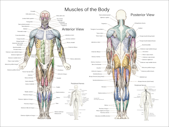
Human Being Anatomy Muscles Anterior View Image Visual Dictionary Online from www.visualdictionaryonline.com There are 20 forearm muscles which are arranged an anterior compartment that contains flexor forearm muscles in the posterior compartment of the forearm are popularly called the extensor the anterior growth causes the vertebral bodies and discs to bulge laterally toward the convexity and to. This quiz requires labeling, so it will test your knowledge on how to identify these muscles (latissimus dorsi, trapezius, deltoid, biceps brachii, triceps brachii, brachioradialis, pectoralis major, serratus anterior, rectus abdominis, etc.). Deep to the gluteus maximus is the gluteus medius, and deep. Labeled anterior and posterior muscles of the body. In the anterior compartment, they are split into three categories: And, together with the scaffolding provided by the skeleton, muscles also determine the form and contours of our body. Flexor carpi ulnaris is the most medial of the superficial flexors. Learn anterior muscles facts using a simple interactive process (flashcard, matching, or multiple choice).
These are the rectus abdominis, pyramidalis, external abdominal oblique, internal abdominal oblique and transversus abdominis.
Standard anatomical terms of location are used to unambiguously describe the anatomy of animals, including humans.the terms, typically derived from latin or greek roots, describe something in its standard anatomical position.this position provides a definition of what is at the front (anterior), behind (posterior) and so on. Anatomynote.com found human body parts labeled anterior view and posterior view from plenty of anatomical pictures on the internet. This is an online quiz called muscles of the anterior surface of the body. The anterior and middle scalene muscles, which also are located at the sides of the neck, act ipsilaterally to rotate the neck, as well as to elevate the first rib. Flexor carpi ulnaris is the most medial of the superficial flexors. Anterior tibialis thick muscle enabling the foot to flex on the leg and to draw near the median axis of the body; Finally a format that helps you memorize and understand. The splenius capitis and splenius cervicis, which are located in the back of the neck, work to rotate the head. In the anterior compartment, they are split into three categories: Muscle tone provides a slight tension on the muscle to prevent damage to the muscle and joints from sudden movements, and also helps to maintain the body's posture. There is a printable worksheet available for download here so you can take the quiz with pen and paper. The muscles found in the anterior compartment of the leg are: There are five muscles that form the abdominal part of the anterior trunk.
Jul 26, 2021 · iliopsoas acts as the antagonist of the gluteus maximus muscle and the hamstring. There are around 650 skeletal muscles within the typical human body. Muscle tone provides a slight tension on the muscle to prevent damage to the muscle and joints from sudden movements, and also helps to maintain the body's posture. Flexor carpi ulnaris is a fusiform muscle located in the anterior compartment of the forearm. Anterior & posterior muscles of the human body vintage muscle anatomy images showing over 50 muscles of anterior and posterior aspect of the human body.
Major Muscles Of The Human Body Youtube from i.ytimg.com We think this is the most useful anatomy picture that you need. As previously mentioned, they are dorsiflexors. This is a table of skeletal muscles of the human anatomy. Learn anterior muscles facts using a simple interactive process (flashcard, matching, or multiple choice). Make writing personal training programs easy with these custom designed exercise templates, and keep your clients focused and progressing. Click on the tags below to find other quizzes on the same subject. Standard anatomical terms of location are used to unambiguously describe the anatomy of animals, including humans.the terms, typically derived from latin or greek roots, describe something in its standard anatomical position.this position provides a definition of what is at the front (anterior), behind (posterior) and so on. This is an online quiz called muscles of the anterior surface of the body.
You can click the image to magnify if you cannot see clearly.
Jul 26, 2021 · iliopsoas acts as the antagonist of the gluteus maximus muscle and the hamstring. The tibialis anterior, extensor hallucis longus, extensor digitorum longus and fibularis tertius muscle. You can click the image to magnify if you cannot see clearly. Deep to the gluteus maximus is the gluteus medius, and deep. Anatomynote.com found human body parts labeled anterior view and posterior view from plenty of anatomical pictures on the internet. There are more than 600 skeletal muscles in the body. These are the rectus abdominis, pyramidalis, external abdominal oblique, internal abdominal oblique and transversus abdominis. The reason for this is their origin at specific points on the tibia or fibula and insertion on certain areas of the foot. This is an online quiz called muscles of the anterior surface of the body. The psoas major and iliacus make up the iliopsoas group.some of the largest and most powerful muscles in the body are the gluteal muscles or gluteal group.the gluteus maximus is the largest; It belongs to the superficial flexors of the forearm, along with pronator teres, palmaris longus, flexor digitorum superficialis and flexor carpi radialis. Deep layers of the torso, upper and lower extremities. Learn anterior muscles facts using a simple interactive process (flashcard, matching, or multiple choice).
We hope you can get the. The posterior tibial allows the foot to extend. The superficial muscles in the anterior compartment are the flexor carpi ulnaris, palmaris longus, flexor carpi radialis. Flexor carpi ulnaris is a fusiform muscle located in the anterior compartment of the forearm. In addition to these, the end of the iliopsoas muscle passes into the anterior compartment.
Muscle Anatomy Posters Anterior Posterior Deep Layers from www.dcfirst.com The splenius capitis and splenius cervicis, which are located in the back of the neck, work to rotate the head. It belongs to the superficial flexors of the forearm, along with pronator teres, palmaris longus, flexor digitorum superficialis and flexor carpi radialis. We hope you can get the. The first three are classified as vertical muscles and they are located near the midline. Finally a format that helps you memorize and understand. The posterior tibial allows the foot to extend. There are around 650 skeletal muscles within the typical human body. There are more than 600 skeletal muscles in the body.
Deep layers of the torso, upper and lower extremities.
We think this is the most useful anatomy picture that you need. Collectively, they constitute 40% to 50% of our body weight. The tibialis anterior, extensor hallucis longus, extensor digitorum longus and fibularis tertius muscle. All muscles maintain some amount of muscle tone at all times, unless the muscle has been disconnected from the central nervous system due to nerve damage. The splenius capitis and splenius cervicis, which are located in the back of the neck, work to rotate the head. Side bending also is an important action of the cervical spine. Almost every muscle constitutes one part of a pair of identical bilateral muscles, found on both sides, resulting in approximately 320 pairs of muscles, as presented in this article. Finally a format that helps you memorize and understand. These are the rectus abdominis, pyramidalis, external abdominal oblique, internal abdominal oblique and transversus abdominis. You can click the image to magnify if you cannot see clearly. Flexor carpi ulnaris is the most medial of the superficial flexors. Gluteal region muscles that move the femur. The psoas major and iliacus make up the iliopsoas group.some of the largest and most powerful muscles in the body are the gluteal muscles or gluteal group.the gluteus maximus is the largest;